Revolutionizing Pathology for Accessible, High-Quality Diagnostics:
Leveraging technology to create digital pathology solutions that enhance the accessibility, efficiency and accuracy of surgical pathology services across healthcare systems.
Let's Work Together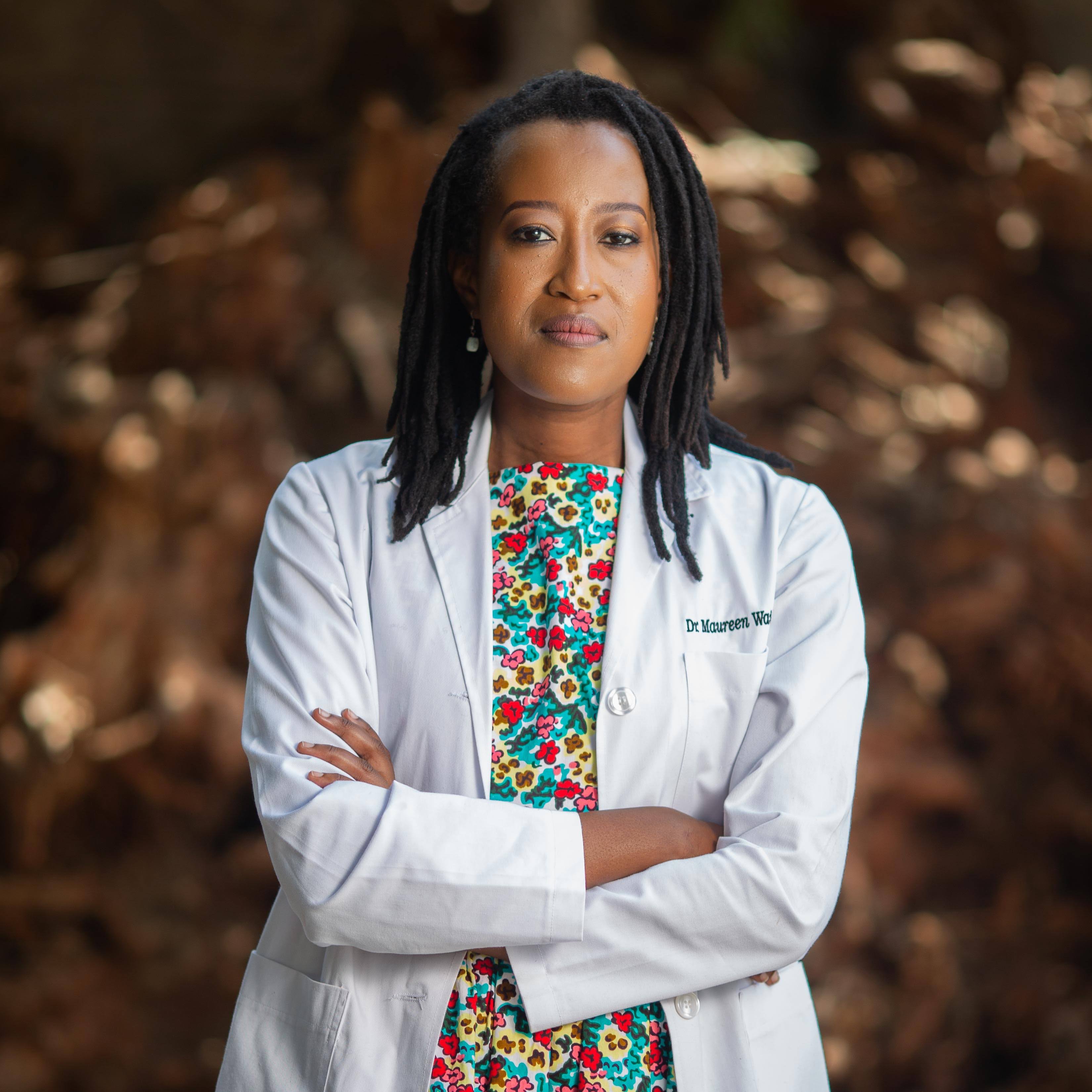
Enhancing Pathology with Digital Technology.
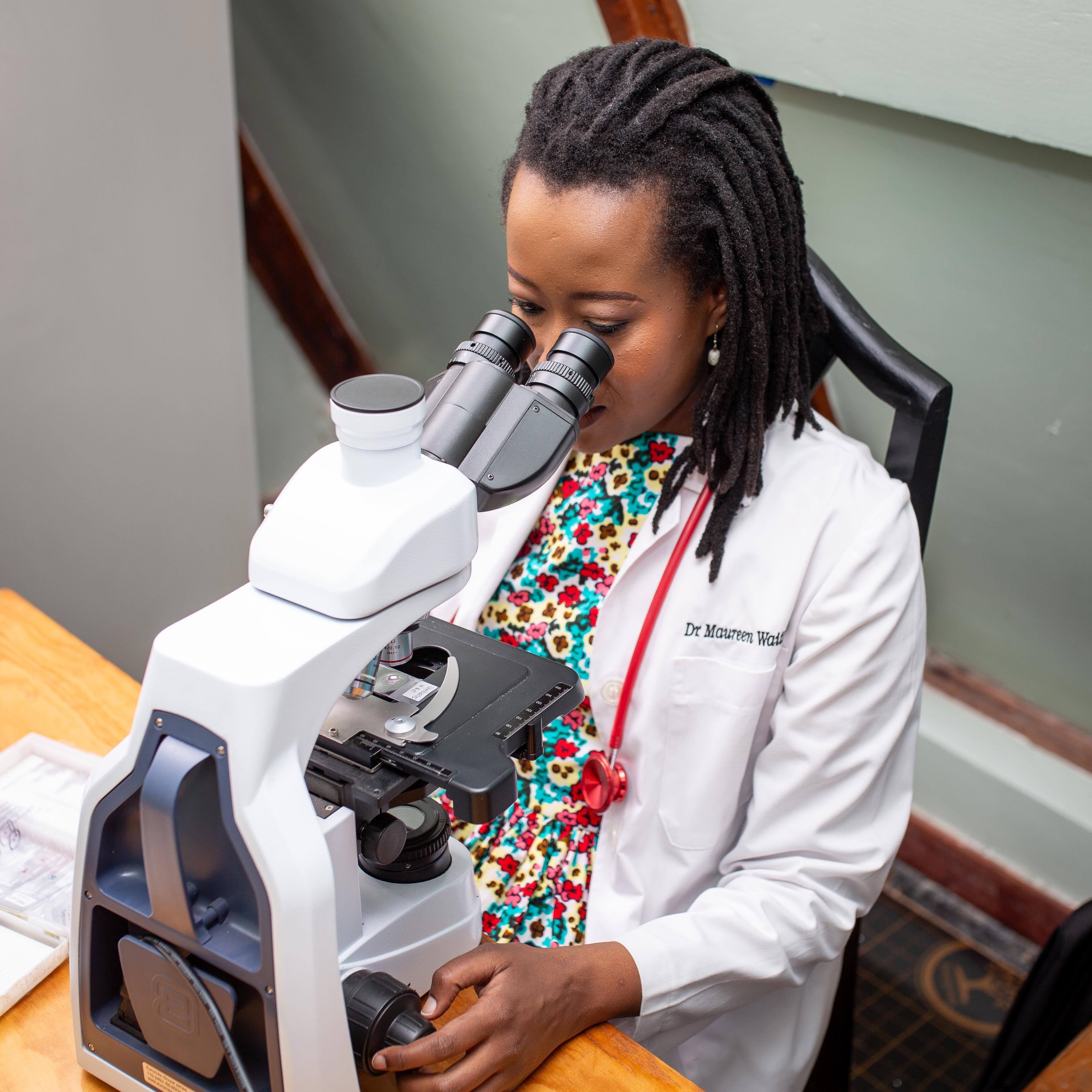
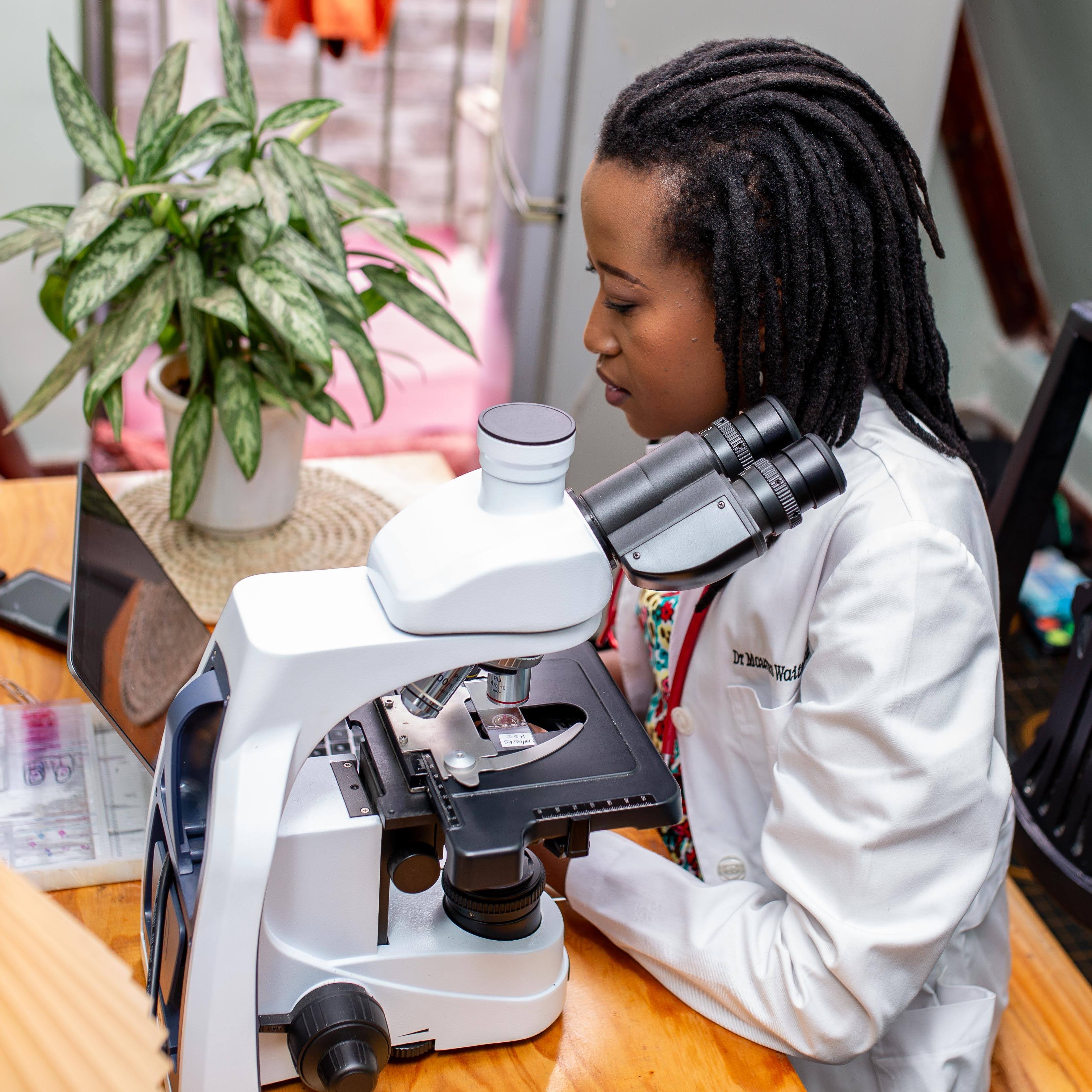
Maureen Waithaka is an experienced medical doctor with specialisation in pathology. She is a passionate medical educator and a digital pathology innovator. She foresees a future where all patients in Kenya, on the continent and beyond, have access to good quality and affordable pathology resources.
An alumnus of Aga Khan University Hospital, a competitive and peer approved anatomic pathology residency program, she has worked both in Kenya and Namibia honing histopathology diagnostic skills.
Learn More About →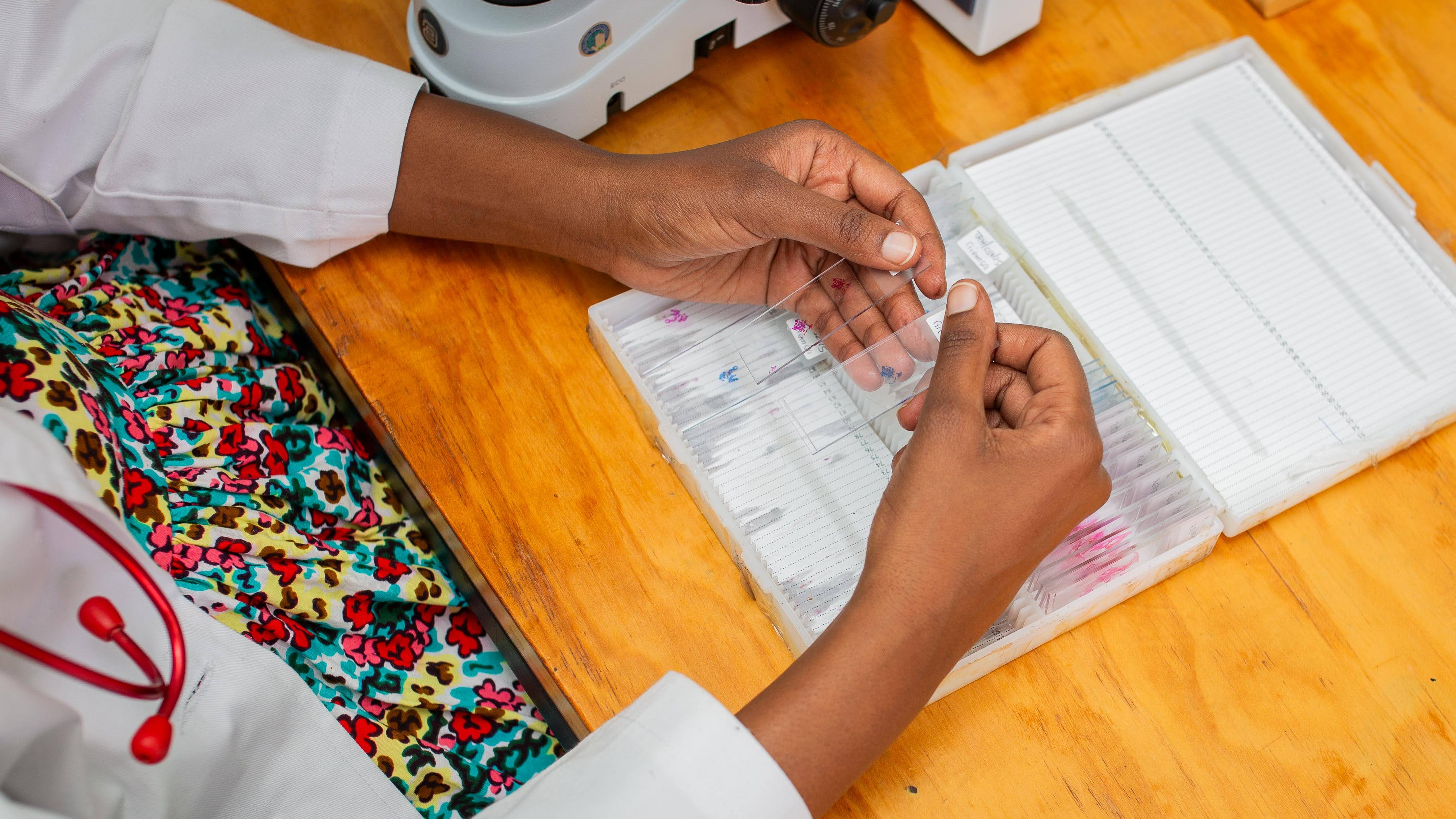
My key areas of expertise.
Histopathology Diagnostics
Developing expertise in the microscopic study of tissue to produce detailed, high caliber and standardized pathology reports that inform disease treatment, including cancer.
Digital Pathology Leadership
Leading implementation of digital pathology solutions and their promising digital pathways that work to improve access to pathology services and patient outcomes.
Medical Education & Content Development
Creating innovative education content for both curricular medical education and public health awareness through digital platforms and traditional teaching methods.
I've worked with top healthcare institutions to enhance diagnostics.
Traditional pathology has long been the mainstay of diagnostics in the institutions within which I have worked. It has laid the foundation for the work I am currently doing, applying digital pathology to revolutionize pathology.
Digital pathology offers a potent potential solution to pathologist shortages faced in many parts of Kenya, Africa and the world at large. Patients in underserved areas can have their biopsies processed into glass slides, which can then be scanned into high resolution digital images that can be securely sent to a pathologist for review and diagnosis, regardless of distance or location. And this is only the tip of the iceberg.
Not only can digital pathology enable remote populations to access crucial pathology services, but it can also make the pathology workflow more efficient. Additionally, digital pathology can integrate AI and molecular data to enable more accurate and more comprehensive diagnoses that improve treatment outcomes.
Get started with a consultation today.
Ensure accurate diagnoses and expert insights with a professional digital pathology consultation. Whether you need diagnostic support, second opinions, or digital pathology solutions, I'm here to help enhance patient care and outcomes.
Let's Work TogetherUnderstanding Pathology Workflows
Explore the evolution from traditional microscopy to modern digital pathology systems
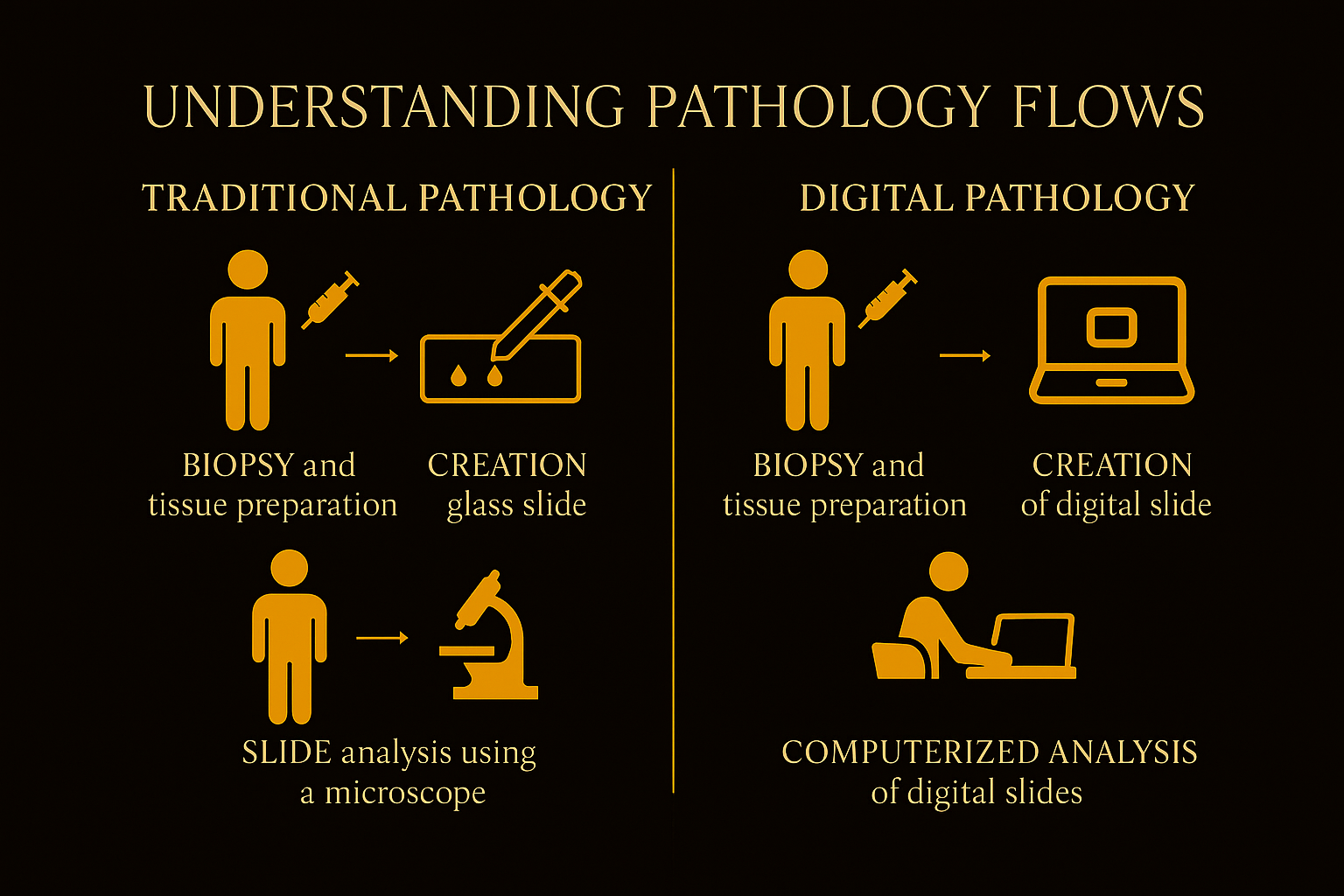
How Traditional Pathology Works
The established workflow that has been the foundation of pathological diagnosis for decades
Biopsy and Tissue Preparation
Under sterile conditions, a doctor takes a biopsy - a sample of tissue from the area of disease, from the patient...
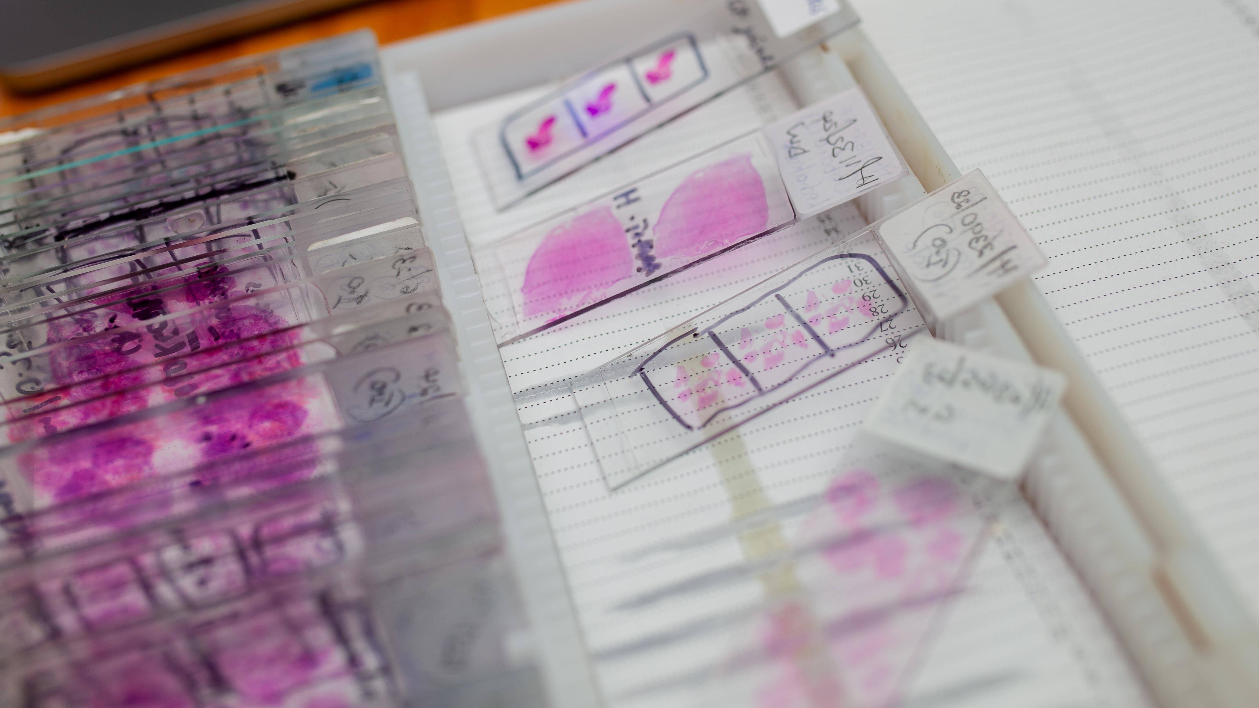
Creation of Glass Slides
In the lab, through a multistep process that involves both manual and automated procedures...
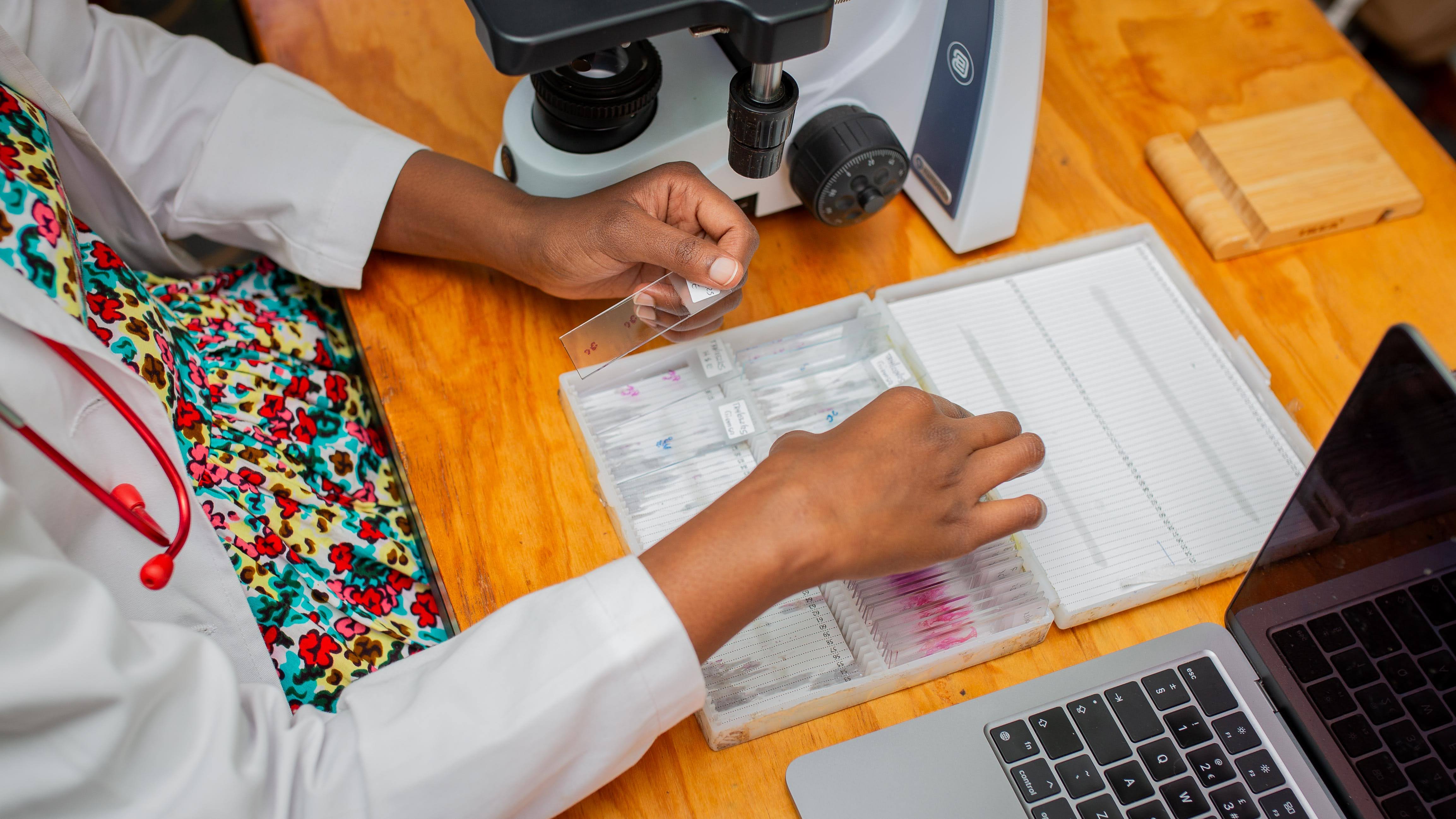
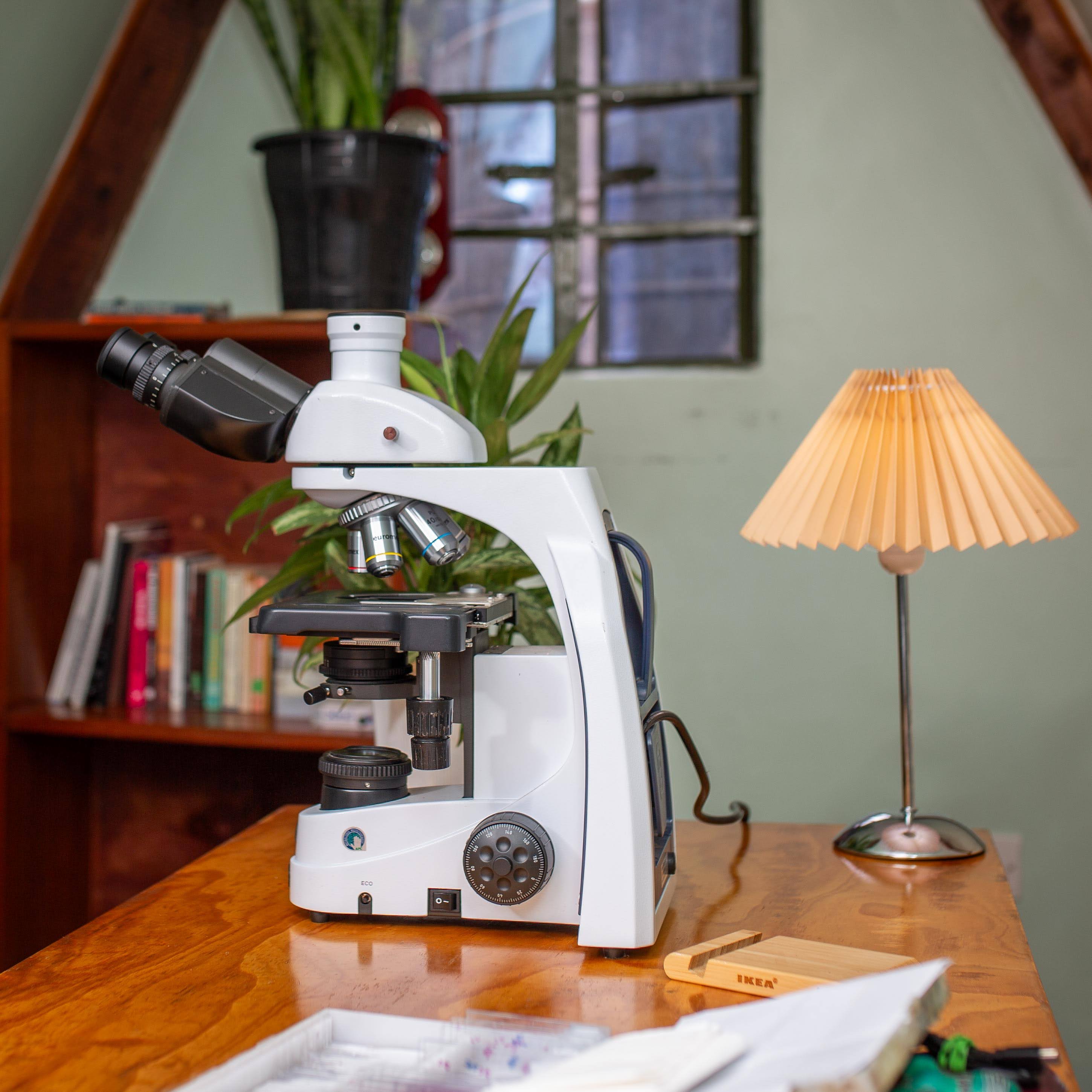
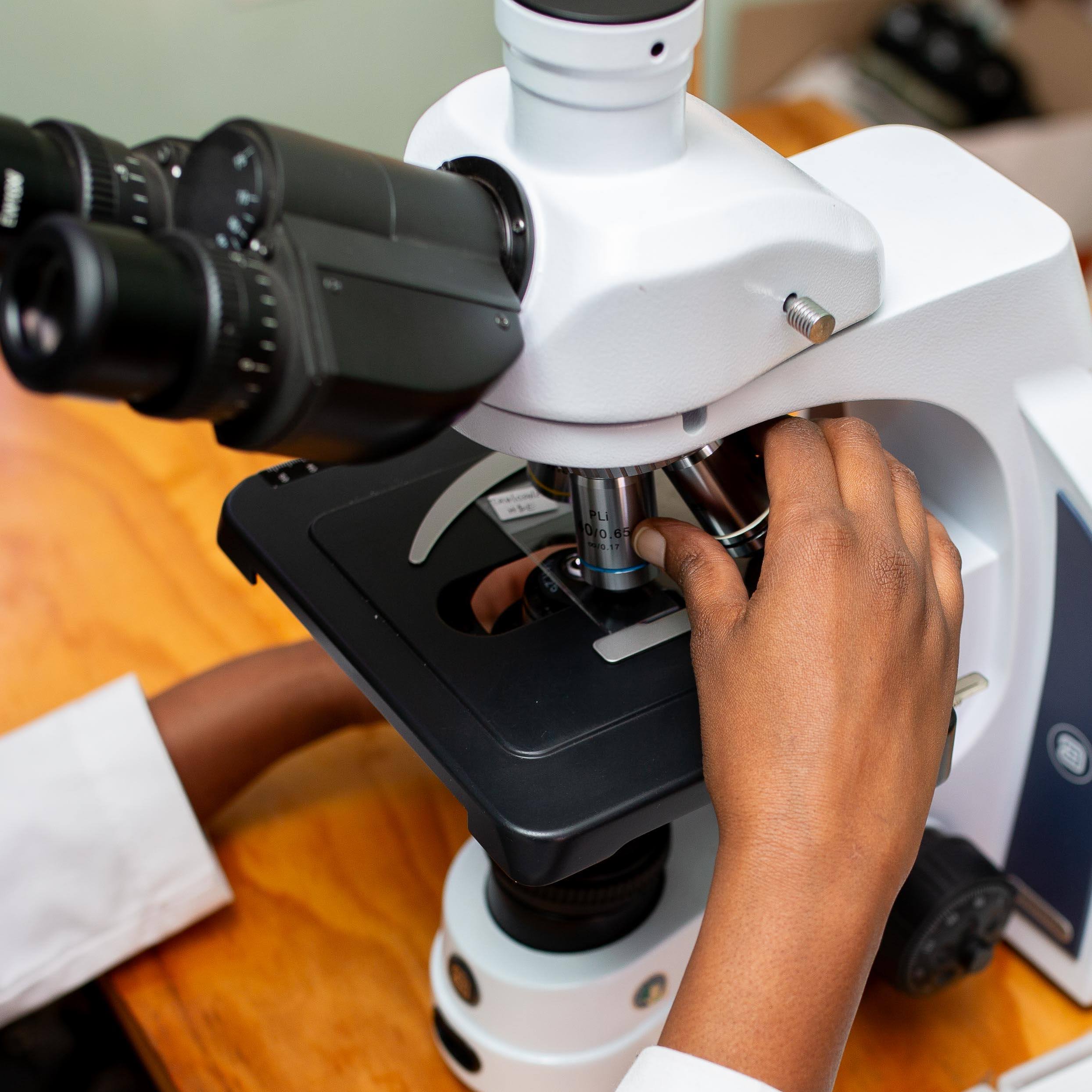
Slide Analysis Using a Microscope
The glass slide is then availed to a pathologist, who examines it under the microscope...
How Digital Pathology Works
The modern approach utilizing digital technology to enhance diagnostic capabilities and workflow efficiency
Biopsy and Tissue Preparation
Under sterile conditions, a doctor takes a biopsy - a sample of tissue from the area of disease...
Creation of Digital Slides
In the lab, through a multistep process, the preserved biopsy is converted into a glass slide...
Computerized Analysis of Digital Slides
The digital slide is examined by a pathologist on the digital platform...